
Of every 100 people
who have an arteriovenous malformation, or AVM, of the brain, about four will experience bleeding in the brain in a given year.
The cumulative risk of bleeding from an AVM over a person’s lifetime can be high, so getting the right treatment is important.
AVMs can develop in the brain, spinal cord or other parts of the body. They may be caused by a problem with the formation of blood vessels in the womb or by an inherited condition.
The exact cause as to why they develop is currently not known.
Symptoms can vary, depending on the location of the AVM in the brain.
Symptoms may begin at any age but usually emerge between ages
10 and 40.
Some AVMs might not cause symptoms at all and are typically discovered in the course of treating another condition.
Other brain AVMs may become so large that they start to push at surrounding tissues and people with AVMs may experience symptoms that are reminiscent of a stroke.
- AVMs can cause:
- weakness,
- or paralysis on one side of the body,
- problems with speech, vision, balance, memory or hearing.

Some people can get headaches, or seizures.
If you are experiencing any such symptoms, you should come to immediate medical attention, and share your health information with a health care provider. A brain imaging study can inform the doctor of where the brain AVM is located, and whether it is bleeding.
As an AVM enlarges or, even worse, begins to bleed, you might experience similar but sudden and severe symptoms.
Usually, you will get a headache and you may pass out. As an AVM ruptures, this can become a life-threatening emergency. A CT scan performed in the ED can quickly and reliably determine if the AVM has started to bleed. The American Stroke Association classifies an AVM bleed as a hemorrhagic stroke. A stroke is an umbrella term for many different types of strokes with a brain hemorrhage due to an AVM being a rare subtype that occurs primarily in younger adults or children.
Brain AVM treatment focuses on eliminating that risk by removing or blocking the abnormal vessels.
There are usually many doctors involved that work together to determine the best course of treatment, depending on the size and location of the AVM, your overall health and any other relevant factors.
Catheter treatment for AVMs
Catheter treatment, or endovascular treatment, is a minimally invasive arteriovenous malformation treatment in which a catheter is inserted through a small incision in the groin or wrist and directed to the site of the AVM in the brain.
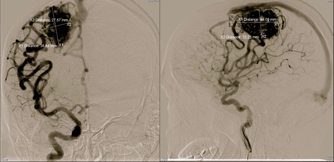
The neurosurgeon will use an angiogram to map out all the blood vessels using dye and x-rays. Once the catheter is in place, an embolizing, or blocking, agent such as a medical glue, or a platinum coil is placed into the AVM to close up the abnormal connections. This is referred to as embolization procedure.
It is performed in a hospital utilizing a specialized X-ray machine under general anesthesia. After the procedure you will be monitored very carefully to see how your brain adjusts to the changes in blood flow that will be occurring in the brain tissue surrounding the AVM. You will be typically released within 24 – 48 hours.
Embolization can be used to completely seal the AVM, or it might be used to reduce the blood flow to a very large AVM before surgery.
Brain surgery for AVMs
Surgery is a highly effective treatment that removes the AVM completely.
The abnormal blood vessels don’t grow back, so surgery can eliminate the risk of future bleeding.
Surgery is performed in a hospital under general anesthesia and requires a craniotomy, a procedure to open the skull so that neurosurgeon can reach the AVM. Each vessel leading to the abnormal tangle is clamped off, and the entire AVM is removed, leaving all brain tissue intact. Depending on circumstances, your surgeons may perform an endovascular embolization procedure a day or so before surgery to reduce blood flow to the AVM.
During the surgery, while you are asleep, an angiogram will be performed to check the surgery site and make sure that all blood vessels were successfully removed.
After surgery, you’ll be monitored in the ICU for a day or so and remain hospitalized for a couple days. Recuperation at home takes about a month.
Although surgery can eliminate an AVM entirely, this procedure may not be the right choice for everyone. Some AVMs are located deep in the brain where surgery becomes risky or impossible, or they may be small enough that they can be resolved with less invasive treatments.
Gamma Knife Radiosurgery for AVMs
Gamma knife radiosurgery is a completely non-surgical arteriovenous malformation treatment for smaller AVMs and those located in areas of the brain where surgery isn’t appropriate.
Noninvasive and completed in less than one hour, radiosurgery delivers a focused dose of gamma radiation directly to the abnormal cluster of vessels.
This burst of radiation damages the walls of the AVM’s blood vessels, causing them to thicken, but that process is slow. It can take two or three years for the AVM to completely close and eliminate the risk of bleeding in the brain. While the AVM is closing, the risk of bleeding still exists, so those who have radiosurgery typically need regular follow-ups for monitoring the status of the AVM.
Although the gamma knife surgery itself may be brief, the mapping of the AVM and preparations may take several hours.
Doctors will need to perform a variety of imaging procedures in order to pinpoint the AVM and determine a treatment plan. Radiation is painless, but you may have side effects such as headaches or nausea after treatment. AVM radiation is typically done only once. There can be also side effects that occur much later in life and are related to radiation injury. For most people, there’s no need for hospitalization after radiosurgery.

About Dr. Dorothea Altschul
Dr. Dorothea Altschul is an accomplished neurointerventionalist in North Jersey and is the Clinical Director of Endovascular Services at Neurosurgeons of New Jersey, practicing out of their Ridgewood office located on East Ridgewood Avenue.
Please call today to schedule a consultation with me. (551) 284-3265
Request a consultation with Dr. Altschul