Do I Need Surgery for Chiari Malformation?
What is Chiari Malformation?
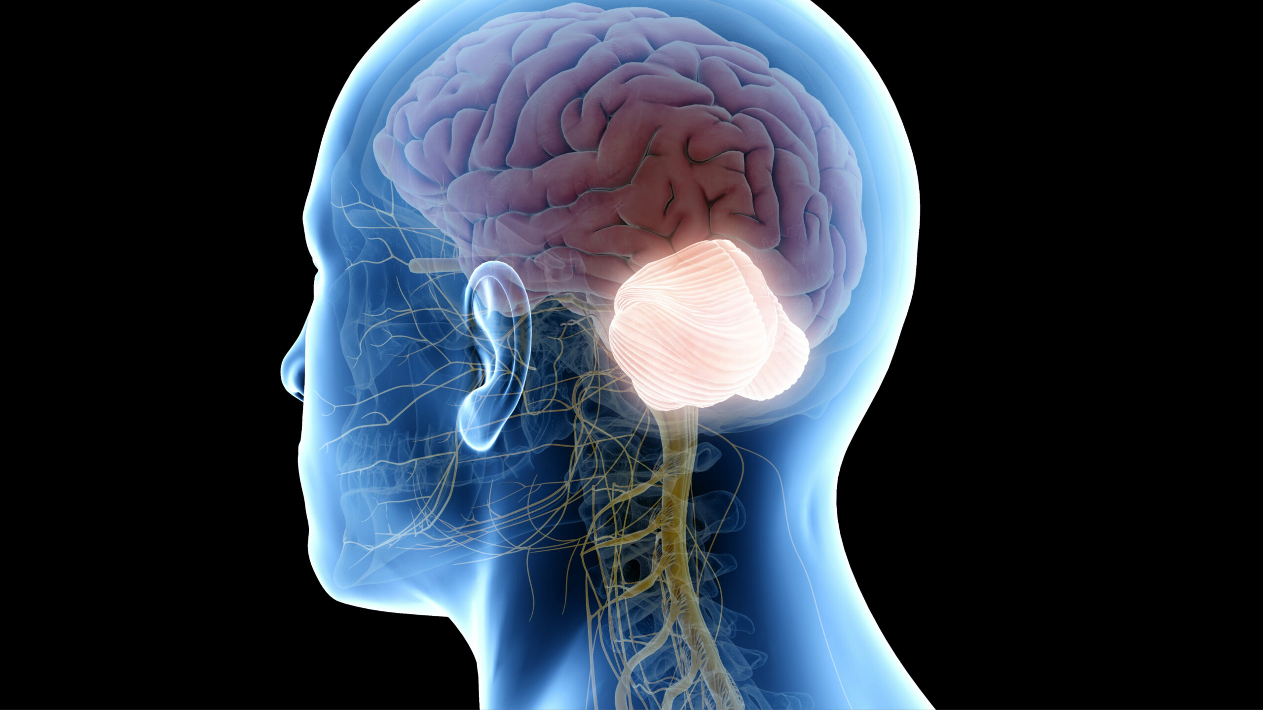
Chiari malformation is a problem in which a part of the brain (the cerebellum) at the back of the skull bulges through a normal opening in the skull where it joins the spinal canal.
This puts pressure on parts of the brain and spinal cord and can cause mild to severe symptoms. In most cases, the problem is present at birth.
For individuals diagnosed with the most common type, Chiari I malformation, one question often arises: When is surgery necessary?
Understanding Chiari Malformation
Chiari malformations affect the area of the brain where the skull meets the spine.
The cerebellar tonsils, located in this region, extend into the spinal canal, potentially compressing important neural structures like the brain stem. This disrupts the flow of cerebrospinal fluid (CSF) that surrounds the brain and spinal cord.
Symptoms can vary greatly between individuals. While many people may have no symptoms, others experience significant discomfort, including neck pain, balance issues, and severe headaches. Straining often worsens these symptoms
Indicators for Surgical Intervention
Most cases of Chiari malformation do not require surgery. Patients without symptoms very rarely require any intervention. Many individuals with Chiari symptoms manage to control them with pain relievers, physical therapy, or lifestyle adjustments.
Some indications that surgery may be necessary are:
- Severe, persistent symptoms that interfere with daily life despite trials of non-surgical therapies.
- Disruption or blockage of spinal fluid which leads to the accumulation of cerebrospinal fluid (CSF)
- The formation of cysts in the spinal cord (syringomyelia, hydromelia, or syrinx)
- Neurological decline such as weakness or numbness in the arms or legs, difficulty swallowing, or issues with eye movement.
- Chronic sleep apnea
Goals of Chiari Decompression Surgery
The primary goal of Chiari decompression surgery is to relieve pressure on the brain stem and spinal cord and restore regular flow of cerebrospinal fluid.
This approach aims to relieve structural compression, improve symptoms, and reverse the progression of cyst formation within the spinal cord.
Navigating the Decision to have Surgery
The decision to proceed with surgery for Chiari malformation often follows an extensive period of monitoring symptoms, conservative treatments, and detailed discussions between the patient and their healthcare providers.
No two cases are identical, and the patient’s lifestyle, goals, and concerns play a major role in decision-making. What works as a treatment for one person might not be as effective for another.
Let's build your treatment
path, together.
What Happens During Cervical Laminoplasty?
The Surgical Procedure
The standard surgical treatment for Chiari malformation is posterior fossa decompression with C1 laminectomy, a procedure performed under general anesthesia by a neurosurgeon.
The surgeon removes a small section of bone at the back of the skull and upper neck. This relieves pressure by giving the brain and spinal cord more room.
In some cases, the surgeon will need to open the dura mater, the membrane covering the brain, to view the tonsils of the cerebellum. If these structures are blocking the flow of cerebrospinal fluid, the surgeon might cauterize or shrink them.
Pre-operative Preparations and Considerations:
Before surgery, patients undergo assessment to ensure they are suitable candidates for the procedure. These evaluations focus on understanding the extent of the malformation, the severity of the symptoms, and how the condition impacts the individual’s daily life.
Critical imaging diagnostic tools include MRI scans of the brain and spine to assess the specific structures involved. Specialized sequences can image cerebrospinal fluid (CSF) flow through the area in question (the cerebellar tonsils and the posterior fossa).
Additionally, patients engage in thorough discussions with their healthcare team about the surgery’s expected outcomes and potential risks.
How is Surgery Performed
Chiari decompression surgery is usually performed under general anesthesia. It is a meticulous procedure that requires several hours.
The patient is positioned face down, and the surgical area is exposed. The surgeon performs a craniectomy which involves removing a small section of the skull to relieve pressure at the site. In most cases, the surgeon also removes a small part of the first spinal bone (laminectomy) to fully decompress the area.
The dura mater (the cover that protects the brain and spinal cord) is opened. The surgeon examines the area below. They want to ensure that nothing is pressing on the cerebellar tonsils. Additionally, they want to make sure that nothing is obstructing the flow of cerebrospinal fluid.
Sometimes, a patch is added to make the dural sac larger, facilitating the free flow of CSF.
Recovery from Surgery
Patients are monitored in a recovery room after surgery. They are then transferred to either a regular hospital room or a neurological/ICU for further observation.
Here’s what patients can generally expect during the recovery process:
- Hospital stay: Most people remain in the hospital for 1-3 days after surgery.
- Pain management: It’s normal to experience some discomfort and neck pain. Pain relief methods vary and can include medication and practical support like using special pillows.
- Activity: After surgery, patients should avoid strenuous activities and heavy lifting for a few weeks to months.
- Gradual recovery: Fatigue is common after the procedure. The body needs time to heal, and patients should increase their activity level slowly.
One crucial aspect in the post-surgery period is monitoring for any signs of complications. You should identify signs of infection, CSF leakage, or neurological changes immediately. Regular follow-up appointments are necessary to monitor the patient’s progress and address any concerns that may arise.
What are the risks of surgery?
Like all surgeries, this procedure comes with risks some of the risks of surgery include:
- Infection
- Cerebrospinal fluid leakage
- Fluid in the brain
- Complications in wound healing
It is important to discuss the risks and benefits with your surgeon to decide if surgery is right for you.
While surgery can reduce symptoms for most people, it may not reverse any nerve injury in the spinal canal that has already occurred.
Potential Need for Repeat Surgery
Repeat surgery for Chiari is uncommon. Reasons for repeat surgery include scar tissue development, addressing problems with cranio-spinal alignment or complication management (CSF leak).
Life After Surgery
After surgery, regular follow-up exams and imaging tests are necessary to assess the surgery’s outcome and the flow of cerebrospinal fluid.
Most patients with severe symptoms notice an improvement in their quality-of-life following surgery. While every patient’s experience is different, routine activities, including work or school, are generally resumed within a few weeks to months.
Ongoing therapy may be beneficial for some individuals, particularly if the Chiari malformation has significantly impaired neurological functions or if there were pre-existing conditions like scoliosis.
Conclusion
Chiari malformation is a complex condition, and the decision to undergo surgery involves evaluating symptoms, anatomical considerations, and an understanding of the potential risks, and lifestyle factors.
If surgery is recommended, understanding the procedure, recovery, and what life post-surgery entails is crucial.
If you are struggling with this condition, contact our office to schedule a consultation with one of our board-certified neurosurgeons who specialize in surgery for Chiari malformation treatment, and let us help you on the path to relief and recovery.

About Dr. Gaetan Moise
Dr. Gaetan Moise is an accomplished neurosurgeon in North Jersey and is a proud member of Neurosurgeons of New Jersey, practicing out of their Ridgewood office conveniently located on East Ridgewood Avenue. His compassionate evidence-based, results-driven approach is guided by his desire to help patients achieve happy, pain-free lives through non-surgical and appropriate surgical solutions. Dr. Moise’s techniques are influenced by the advancements in minimally invasive surgery technology as well as advances in the understanding of the intricacies of the nervous system, brain, and spinal cord. Dr. Moise is a member of The Congress of Neurological Surgeons and the American Association of Neurological Surgeons. He is accepting new patients.