The brain is located in a closed space surrounded by the skull. Underneath the skull is type of covering of the brain, called the dura that surrounds the brain. This layer keeps the ridges of the skull smooth so the brain is better protected. Underneath the dura is yet another thin layer called the arachnoid which layers very carefully around all the nooks and crannies of the brain. Sustaining head injuries can cause many different types of bleeding inside the brain depending on the type of injury, and whether it is a serious or minor head injury.
A subdural hematoma is just one specific kind of a bleeding.
It can be the only bleeding or it can come with other bleeding types.
In an acute subdural hematoma, bleeding occurs between the dura and the arachnoid on the surface of the brain, but not actually in it. As bleeding continues it may start causing pressure on the brain by pushing at the brain tissue, and that can cause symptoms.
Symptoms depend on the rate of bleeding.
Fast growing hematomas caused by tearing of tiny blood vessels on the surface, can become a life-threatening emergency.
Younger adults can pass out immediately, or even become comatose, requiring emergency treatment.
Slow-growing hematomas can go on for days or weeks before they can cause symptoms.
You may experience
- unusual headaches,
- confusion,
- dizziness,
- or trouble speaking along with trouble walking or falls.
Some people may not experience any symptoms at all, but may get seizures. How you respond to the subdural hematoma depends not only on the size of the hematoma but also on your state of health and age.
If you have a bleeding disorder or take blood thinners to treat a medical condition, you maybe at higher risk for a subdural hematoma.
A rapidly growing hematoma in the subdural space is a collection of very acute and fresh blood and it will typically require surgical treatment.
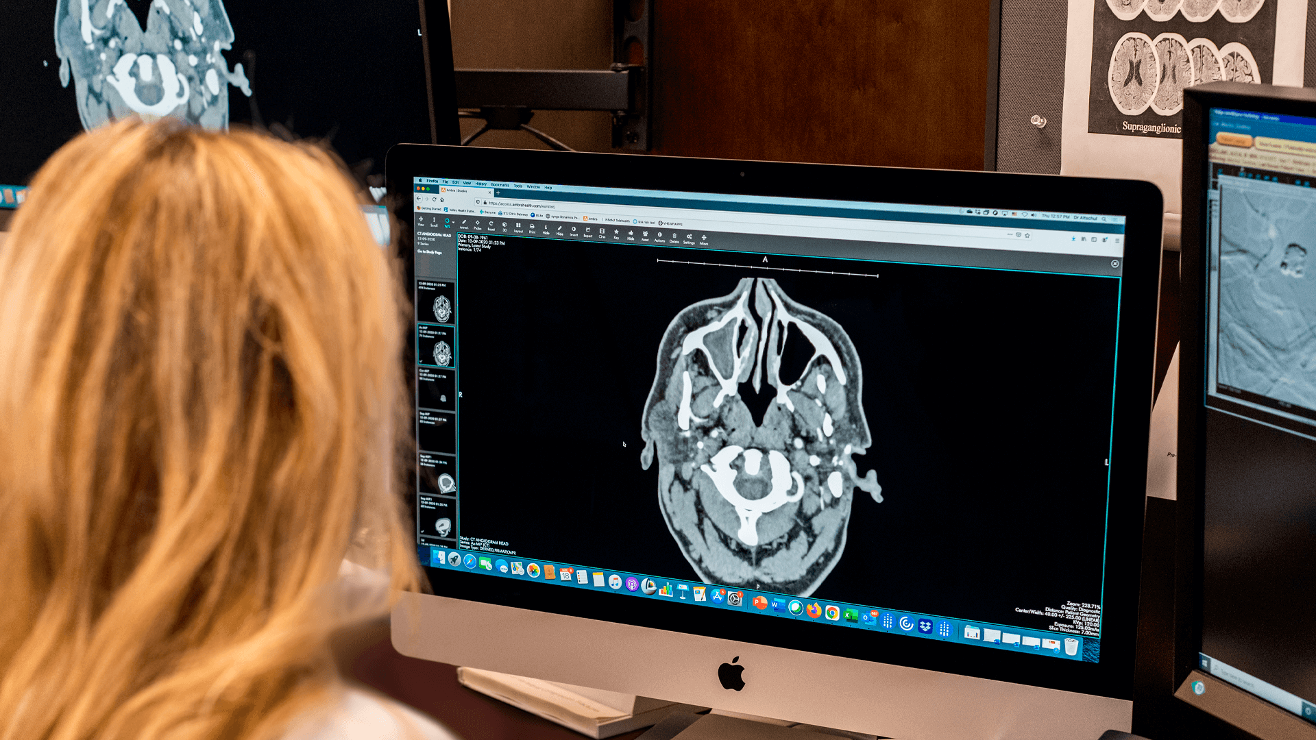
A CT scan can diagnose the subdural hematoma. That scan usually gets done in the emergency room, and a neurosurgeon will perform a small hole in the skull to drain the blood out. Sometimes a larger opening, called a craniotomy, is necessary to treat subdural blood. If a person has a bleeding problem or is taking blood thinners , measures will be taken to improve blood clotting. This may include giving medicines or blood products, and reversal of any blood thinners, when possible. Other medications to help reduce swelling or pressure in the brain or control seizures may also be used.
Slow-growing hematomas are more common in older people, particularly elderly men and individuals with a history of alcohol abuse are at risk for a slower kind of bleeding, called a chronic subdural hematoma.
In older adults, the brain has usually shrunken a bit, which creates more space between the dura and the brain. As a result, blood collects slowly over the brain leading to a chronic subdural hematoma.
A CT scan (computed tomography ct) or MRI (magnetic resonance imaging mri) scan can inform the doctor if the bleeding is an acute or chronic type, or sometimes a mixture of both.
Different stages of blood can be identified on those images, and for those reasons any subdural that is not acute, is sometimes referred to as non-acute subdural hematoma. Individuals with non-acute hematomas may not even recall any head trauma.
If you have very few symptoms, your own general practitioner may order a scan to be done in an outpatient setting, and it may come as a surprise to you that you have been able to perform your daily activities with such a bleeding in your head, and have not experienced more symptoms.
The treatment of chronic subdural hematoma depends on their severity.
Treatment for a subdural hematoma include a range of options that go from watchful waiting to requiring brain surgery with one or two burr holes.
Burr holes are quarter sized small holes above your ear done in an operating room by a neurosurgeon. The small hole allows for the blood to get drained out and relieve the high pressure it has caused on the brain. The brain will then slowly expand back to its original position. The brain doesn’t necessarily “spring” back in place particularly if the bleeding has been going on for a while. A small cavity can remain and occasional that can lead to a collection of air, or bloody-type of fluid which fills in that space.
In fact, despite initial surgical and medical treatment, non acute subdural hematomas are notorious for recurrence.
To decrease the chance of that recurrence, your neurosurgeon may offer you an angiogram.
During an angiogram (angiography), a catheter is inserted through an artery in the groin or wrist, and threaded into the arteries of the neck and brain. Special dye is then injected, and an X-ray screen shows blood flow through the arteries and veins. This procedure allows direct access to the main blood supply of the dura, the middle meningeal artery (MMA), and is capable of occluding those blood vessels, and thereby allowing the collection to be resorbed without recurrence.1 In one study, at three month follow up, those individuals who received a MMA treatment were noted to have faster brain re-expansion (34 days versus 98 days) and decreased rate of hematoma recurrence (3.8% versus 33.3%).2 While this treatment method has been known for more than 20 years, it still remains an ongoing area of research. Here at Neurosurgeons of NJ we do evaluate our patients for this type of treatment.
In summary, chronic subdural hematoma is a common condition amongst those aged 65 years or older, and is one of the most common neurosurgical conditions.
Treatment can vary, and you can get more advice on this condition from one of our specialists.

Further reading:
- Kim E. Embolization Therapy for Refractory Hemorrhage in Patients with Chronic Subdural Hematomas. World Neurosurg. 2017;101:520-527.
- https://evtoday.com/articles/2021-feb/middle-meningeal-artery-embolization-for-subdural-hematoma-what-weve-learned-what-we-need-to-know

About Dr. Dorothea Altschul
Dr. Dorothea Altschul is an accomplished neurointerventionalist in North Jersey and is the Clinical Director of Endovascular Services at Neurosurgeons of New Jersey, practicing out of their Ridgewood office located on East Ridgewood Avenue.
Please call today to schedule a consultation with me.
(551) 284-3265
Request a consultation with Dr. Altschul