
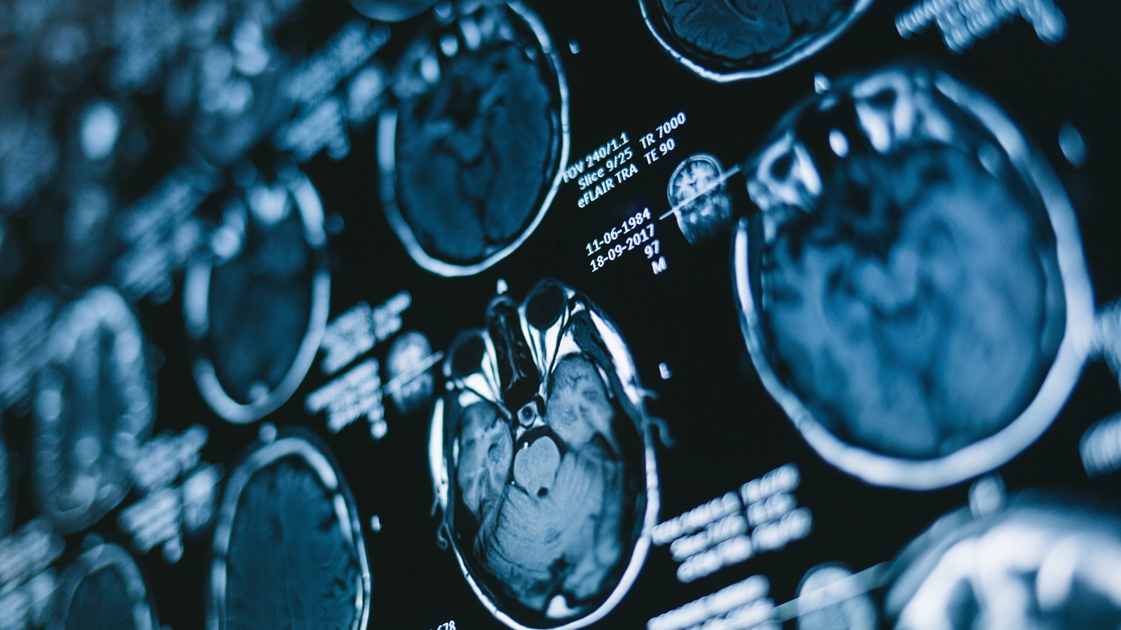

An eye twitch that keeps coming back or spreads to your cheek and mouth can leave you wondering what’s causing it. If your doctor suspects hemifacial spasm, a specialized MRI can help them look closely at your facial nerve and the blood vessels around it. Understanding why the scan is ordered can make the next step feel more manageable.
A hemifacial spasm MRI provides detailed images, but it doesn’t diagnose the condition on its own. Your doctor considers your symptoms, examination and scan together. If you have been diagnosed with hemifacial spasm, do not let a normal MRI report end your evaluation. Have the actual images reviewed by a neurosurgeon who regularly treats hemifacial spasm and performs microvascular decompression (MVD). This review is an essential step before deciding that blood vessel pressure is absent or that an MVD consultation would not help.
Why Doctors Order an MRI for Hemifacial Spasm
Hemifacial spasm causes muscle contractions you can’t control on one side of your face. It often starts as twitching around one eye and may spread to the cheek or mouth over time. According to the National Institute of Neurological Disorders and Stroke, the condition most often develops when a nearby blood vessel presses on the facial nerve, which controls facial movement.
Doctors usually diagnose hemifacial spasm by asking about your symptoms and examining your facial movements. A 2025 review in the Journal of Neurology notes that it remains a clinical diagnosis, but that detailed MRI adds valuable information. The scan helps look for a possible cause and checks the surrounding area. It can also help your surgeon plan treatment if surgery is being considered.
Hemifacial Spasm vs. a Common Eye Twitch
Many people have a harmless eyelid twitch at some point, often linked to stress, fatigue or caffeine. These twitches usually come and go and fade on their own within days or weeks. Hemifacial spasm tends to persist, stays on the same side of the face and can spread beyond the eye to the cheek and mouth. It may even continue during sleep. If your twitching lasts for weeks or begins to involve other facial muscles, it’s worth having it evaluated.
What the MRI Looks For
A Blood Vessel Pressing on the Facial Nerve
Your doctor looks closely at the facial nerve near the brainstem, where it leaves the brain. Pressure from a nearby vessel can irritate the nerve and trigger spasms. Detailed images help show the relationship between these small structures.
A report may describe this as “neurovascular contact” or “neurovascular compression.” These findings need context. A vessel can touch the nerve without causing symptoms, so your specialist checks whether the location and degree of contact fit your facial movements.
Other Possible Causes
MRI also checks for less common causes, such as a growth near the nerve or an abnormal group of blood vessels. It may show changes associated with conditions such as multiple sclerosis. Finding one of these causes can change the treatment plan, but an MRI order alone doesn’t mean your doctor suspects a serious condition.
What Makes a Hemifacial Spasm MRI Different
The facial nerve and nearby vessels are small, so a routine brain MRI may not show every detail. Your doctor may request thin, high-resolution images, sometimes called FIESTA or CISS images. These help the team examine the nerve and surrounding fluid spaces more closely.
Your scan may also include MRA, a type of MRI that shows blood vessels, or images taken with contrast material. The imaging team chooses the types of images based on the questions your doctor needs answered, so not every patient needs the same combination. If you’ve already had a standard brain MRI, ask whether a more detailed scan of the facial nerve area would be helpful.
Are you Ready to Learn More
about Hemifacial Spasm?
What to Expect During Your MRI
Before your appointment, tell the imaging team about implanted devices, metal in your body, kidney disease, previous reactions to MRI contrast and any possibility of pregnancy. Follow the center’s instructions about clothing, jewelry, food and medications. If enclosed spaces make you anxious, let the team know ahead of time so they can discuss ways to help.
During the scan, you’ll lie on a table that moves into the scanner. The machine makes loud tapping or knocking sounds, and the team provides hearing protection. Staying still helps produce clear images, so tell the technologist if spasms make that difficult. You can communicate with them throughout the test.
MRI doesn’t use X-rays, and taking the images shouldn’t hurt. If contrast is needed, you’ll receive it through an IV. Ask the imaging center how long your appointment will take and when your doctor should receive the report.
Understanding Your MRI Results
When the Scan Shows Nerve Compression
Visible compression can help explain your symptoms and guide a discussion about treatment. It doesn’t automatically mean you need surgery. Your specialist reviews the images alongside your examination and asks how the spasms affect activities such as reading, driving or talking with others.
When Your MRI Report Says Normal
A normal MRI report does not rule out hemifacial spasm or automatically rule out MVD as a treatment option. If you have been diagnosed with hemifacial spasm, or your facial movements strongly suggest it, seek an expert review of your actual MRI images. Do not stop at the written report or assume that a surgical consultation is unnecessary.
There are two reasons a scan may be reported as normal even when a blood vessel is pressing on the facial nerve. First, a routine brain MRI may not include the thin, high-resolution images needed to see this small area clearly. Second, the right images may be available, but subtle pressure on the nerve may be overlooked when they are read. These are different problems, and an experienced MVD surgeon can assess both.
The surgeon should review the facial nerve and nearby blood vessels on the images themselves, alongside your symptoms and examination. If the scan lacks the right sequences, the surgeon may order a focused MRI with high-resolution images such as FIESTA or CISS, sometimes with MRA to show the vessels. If those images are already available, a careful second review may identify a finding that the original report did not describe.
The experience of the person reviewing the scan matters. Even a scan read by a skilled radiologist or a surgeon who treats hemifacial spasm less often may need review by a neurosurgeon with specific MVD experience. The question is whether a vessel is pressing on the facial nerve in a way that fits your symptoms. A vessel touching the nerve does not, by itself, prove that it is causing the spasms.
Before accepting that no blood vessel pressure is present, arrange a consultation with an experienced hemifacial spasm surgeon. Ask the imaging center to transfer the full MRI study, including all available sequences, and send the written report as well. Let the surgeon know how the spasms started, which side they affect and whether they have spread beyond the eye.
An expert review cannot guarantee that compression will be found or that surgery is right for you. It helps ensure that a normal report does not prevent a full evaluation of your options. The surgeon can explain whether more imaging is needed and whether MVD or another treatment fits your situation. A consultation does not commit you to surgery.
How MRI Results Help Guide Treatment
Your MRI is one part of choosing treatment. Your symptoms, overall health, response to previous care and preferences also matter. The goal is to find an approach that offers meaningful relief with risks you understand.
Botox Injections
Botulinum toxin (Botox) injections can temporarily reduce spasms by relaxing the affected muscles. Repeat treatments are usually needed because the effects wear off over time. The injections treat the muscle contractions but don’t remove pressure on the nerve.
Microvascular Decompression
Microvascular decompression, or MVD, is surgery to relieve pressure from a blood vessel on the facial nerve. The surgeon carefully separates the vessel from the nerve, often placing a small cushion to keep them apart. For suitable patients, it can provide lasting relief, although results vary and improvement may take time.
Why Expert MRI Review Matters
If you have hemifacial spasm, expert review of your MRI is an essential part of evaluating your treatment options. A normal report alone should not be used to rule out blood vessel pressure or dismiss an MVD consultation. Have the actual images reviewed by a neurosurgeon who regularly treats hemifacial spasm and performs MVD, even if the original report found no compression. The surgeon can check for subtle findings, determine whether the right sequences were obtained and order additional imaging when needed.
Have Your MRI Reviewed by a Hemifacial Spasm Specialist
If you have been diagnosed with hemifacial spasm or have persistent spasms on one side of your face, schedule an expert review of your symptoms and actual MRI images, even if your report says the scan is normal. Do not let that report be the reason you miss an MVD consultation. At Neurosurgeons of New Jersey, Dr. Anthony D’Ambrosio is a fellowship-trained skull base neurosurgeon specializing in hemifacial spasm treatment. He can review your images, assess whether the appropriate MRI sequences were obtained and discuss whether further imaging or an MVD evaluation is appropriate.
Request a consultation with Dr. D’Ambrosio or call 551-284-3265 to schedule an appointment. Ask how to transfer your MRI images and report before your visit, and whether a telehealth consultation is available for you.
Hemifacial Spasm MRI FAQs
Can an MRI diagnose hemifacial spasm?
An MRI can’t diagnose hemifacial spasm by itself. Doctors usually make the diagnosis based on your symptoms and a neurological exam, especially the pattern of twitching that starts near one eye and spreads to the cheek or mouth. The MRI adds important information by looking for a blood vessel pressing on the facial nerve and checking for less common causes, such as a growth near the nerve.
Can I have hemifacial spasm with a normal MRI?
Yes. A normal MRI report does not rule out hemifacial spasm or automatically rule out MVD. If you have been diagnosed with hemifacial spasm, have the actual images reviewed by a neurosurgeon who regularly treats it and performs MVD. Subtle compression can be overlooked, and routine scans may lack the detailed FIESTA or CISS images needed to assess the nerve. Expert review helps determine whether additional imaging is needed and whether an MVD consultation should move forward.
Can a regular brain MRI show hemifacial spasm?
A routine brain MRI can rule out some causes, such as a tumor, but it may not show the facial nerve and nearby blood vessels in enough detail. That’s why doctors often request thin, high-resolution images of the area where the facial nerve leaves the brainstem, sometimes along with MRA. If your first scan was a standard brain MRI, your specialist may recommend a more focused study.
Do I need contrast for a hemifacial spasm MRI?
Not always. Some scans include contrast to help examine certain tissues and look for less common causes, while others rely on detailed images without it. Your doctor and the imaging team decide whether contrast is needed for your scan. Before your appointment, tell them about kidney problems, previous contrast reactions or any possibility of pregnancy so they can plan safely.

About Dr. Anthony D'Ambrosio
Dr. Anthony D'Ambrosio is an accomplished neurosurgeon in North Jersey and a proud member of Neurosurgeons of New Jersey practicing primarily out of their Ridgewood office conveniently located on East Ridgewood Avenue. Dr. D’Ambrosio focuses his clinical practice on brain tumors, nervous system disorders, and facial pain disorders. He has expertise in a variety of complex surgical and radiosurgical techniques as well as minimally invasive procedures intended to successfully treat complex diseases of the brain. These techniques include micro-neurosurgery, microvascular decompression surgery and Gamma Knife radiosurgery. He's authored over 25 peer-reviewed journals and is the recipient of many awards.
Recent Posts:
Hemifacial Spasm MRI: What It Shows and What Results Mean


Please call today to schedule a consultation with me.
(551) 284-3265
Request a consultation with Dr. D'Ambrosio