
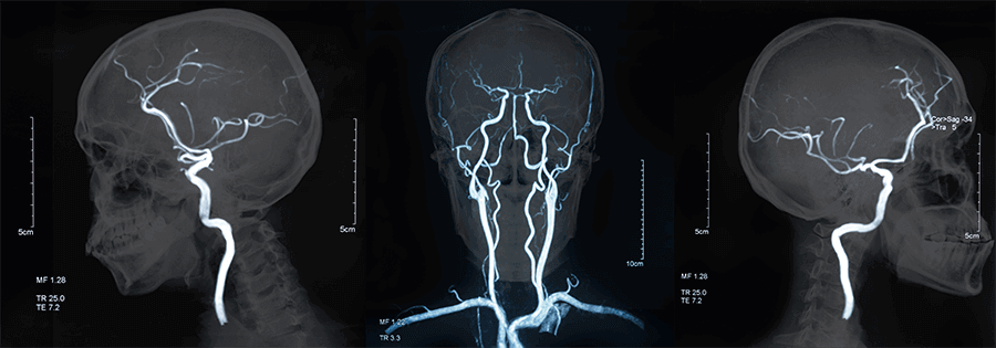
What is a brain angiogram?
An angiogram is a procedure that allows a neurosurgeon to visualize your arteries and veins of the brain.
This is accomplished by injecting a contrast dye into your bloodstream, then taking a specialized X-ray. The dye makes the blood vessels appear on a screen, providing an X- ray image of your blood vessels that could not otherwise be seen.
The difference between an angiogram and other imaging tests, is that the angiogram provides information of the speed of blood flow and visualizes smaller blood vessels than other tests do.
It is more closely related to a video rather than a photograph. It provides information on any type of problem on the blood vessel in the brain, but typically does not show the brain itself. It can be used to visualized brain and spine blood vessels in the neck.
It requires specialized equipment, and hence it is performed in a hospital that has the capability to do this kind of procedure. What can be a bit confusing is the name of the test.
A brain angiogram or cerebral angiogram or cerebral angiography are just different names for the same test.
Angiograms are used to identify and/or define:
- Brain aneurysms,
- brain arteriovenous malformation,
- carotid stenosis and other narrowings of the brain vessels,
- arteriovenous fistulas and other vascular abnormalities,
- blockages in the brain, and also a blood clot such as in a cerebral sinus thrombosis, and other problems with the blood vessels.
How is the brain angiogram performed?
The actual procedure takes approximately 20-30 minutes. You will have to come to the hospital on an empty stomach because you will get sedation and pain medication during the procedure to keep you comfortable.
Based on your medical history, current medications and other factors, your neurosurgeon and anesthesiologist will determine what kind of medication you can receive for sedation. You will be also asked if you ever had any allergic reaction to medications or medical dyes, and if you were taking any blood thinner.
Then, you will be taken to the procedural room, where a doctor or nurse will start an IV before the procedure begins. Stickers for the heart monitor will be placed on your chest, and a blood pressure cuff will be placed on both your arms unless there are restrictions applicable to your individual condition.
You will be put on a narrow X-ray table, and you may notice that the room feels cold. You will be given warming blankets to feel more comfortable. You will see your neurosurgeon, nurses, scrub techs, X-ray techs and an anesthesiologist.
You may see them all come together for a “time-out” huddle before the procedure and may even overhear them talking about you.
This is nothing to be alarmed over. In fact, it is a standard safety measure and gives all of the staff a chance to review your pertinent medical history and the details of the procedure out loud to ensure everyone is well informed and on the same page.
The neurosurgeon will identify an access artery using an ultrasound, and then will give you a local anesthetic to numb the area over the artery. The tube called a catheter is inserted through the femoral artery in the groin area in the upper thigh but a brain angiogram can nowadays be performed through the wrist artery as well. You should ask your neurosurgeon which artery she or he is planning to access.
Contrary to common belief, the catheter never enters into your actual brain inside the skull during an angiogram. An angiogram is purely a fact-finding diagnostic test, and will always be needed to first diagnose and characterize a medical condition.
There is a difference between diagnosis and treatment. Once the diagnosis is made, a treatment plan will be determined. Sometimes, treatment is pursued at the same time, and sometime it is scheduled for a later date. You should ask your neurosurgeon to be very specific about that.
An angiogram will typically evaluate your entire brain vessels, front and back, left and right.
Depending on your condition, the neurosurgeon will spend about 10-15 minutes on each side. The catheter can be repositioned through the aorta in the chest on each side. The total procedure time, preparation and immediate aftercare may take altogether one to two hours, during which your loves ones won’t be able to communicate with you.

About Dr. Dorothea Altschul
Dr. Dorothea Altschul is an accomplished neurointerventionalist in North Jersey and is the Clinical Director of Endovascular Services at Neurosurgeons of New Jersey, practicing out of their Ridgewood office located on East Ridgewood Avenue.
Recent Posts:


Please call today to schedule a consultation with me.
(551) 284-3265
Request a consultation with Dr. Altschul