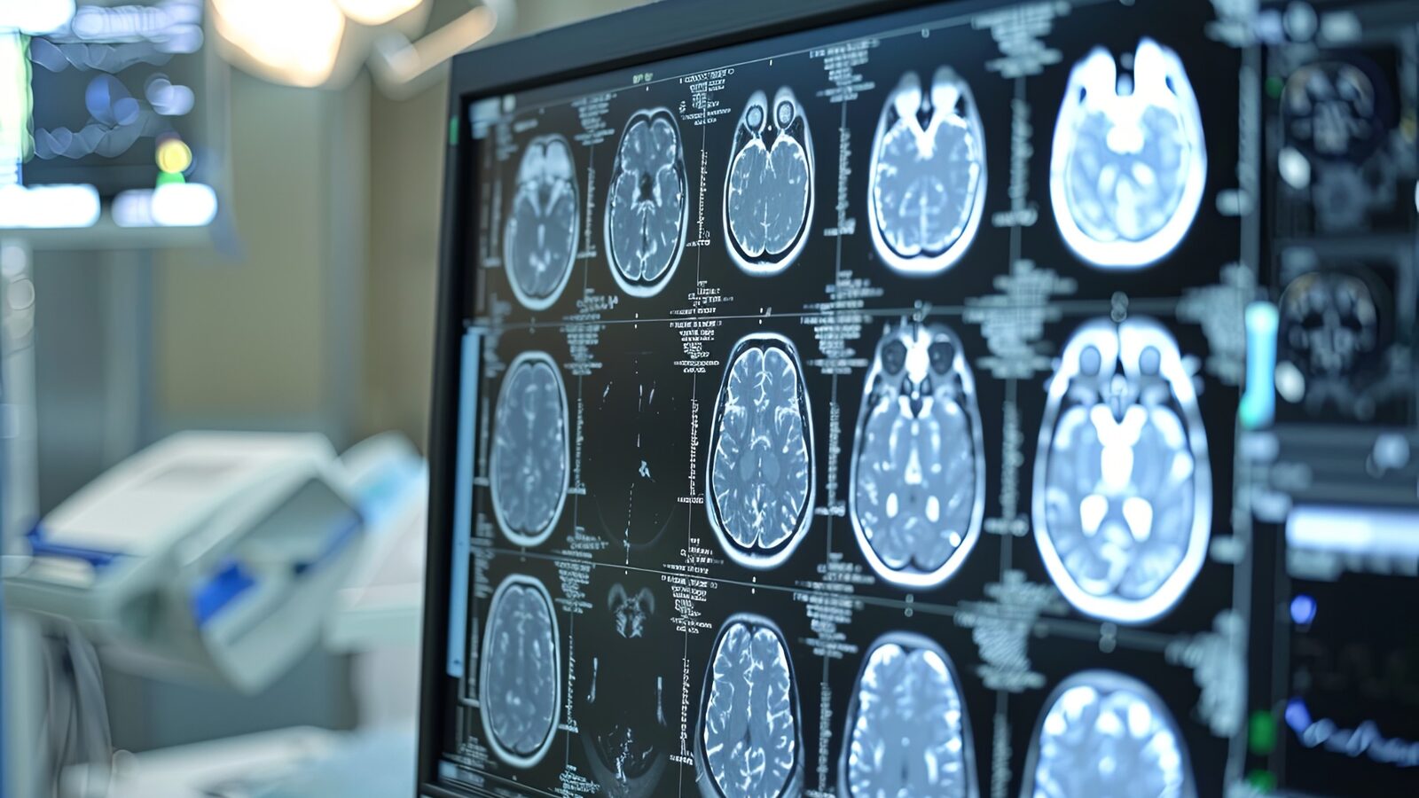

Finding a brain aneurysm on an MRI or CT scan does not automatically mean you need emergency surgery. A neurovascular specialist explains what doctors evaluate next, how they assess rupture risk, and why some aneurysms are monitored while others are treated.
Learning that a brain scan revealed an aneurysm can be alarming, especially when the scan was ordered for an unrelated reason. But an unruptured brain aneurysm is not automatically an emergency. Finding one also does not necessarily mean brain aneurysm surgery is needed.
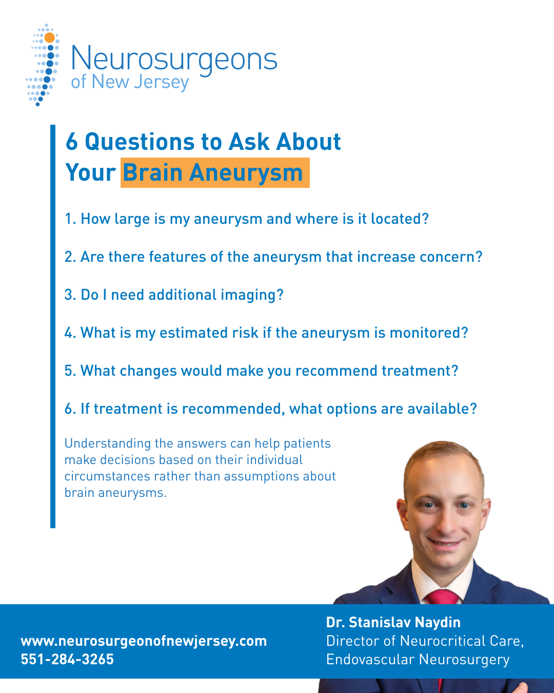
For many patients, the word “aneurysm” sounds like a ticking time bomb. Dr. Stanislav Naydin, Director of Neurocritical Care and Endovascular Neurosurgery at Neurosurgeons of New Jersey, says that is one of the biggest misconceptions about an incidental aneurysm finding.
“Monitoring is an active medical decision, not neglect. It means we’ve weighed the risk of the aneurysm against the risk of treating it, and right now, watching closely is the safer path.” – Dr. Stanislav Naydin
What happens next depends on both the aneurysm and the patient. Doctors consider its size, shape, location, and whether it changes over time. They also consider a patient’s medical history and other risk factors.
Step 1: Have the Scan Reviewed by a Cerebrovascular Specialist
The first step is confirming and understanding the finding.
A brain aneurysm, also called a cerebral aneurysm, is a weakened area in the wall of an artery that bulges outward. Aneurysms can vary in size, shape, location, and risk.
A patient may be referred to a cerebrovascular or neurointerventional specialist who treats diseases of the brain’s blood vessels.
The specialist can review the original scan, confirm the finding, and decide whether more detailed imaging is needed.
The discovery alone does not determine treatment. Some aneurysms require intervention. Others can be monitored over time.
Step 2: Determine Whether More Imaging Is Needed
An MRI or CT scan may be where an aneurysm is first noticed. Doctors may then need a closer look at the brain’s blood vessels.
Additional tests may include a CTA, MRA, or cerebral angiogram. These studies can help doctors determine:
- Where the aneurysm is located
- How large it is
- Its shape
- How it relates to nearby blood vessels
- Whether it has features that could affect treatment
The goal is to understand the aneurysm well enough to decide what should happen next.
Step 3: Evaluate the Risk of Rupture
One of the most important questions is: How likely is this aneurysm to rupture? There is no single answer that applies to every patient.
Dr. Naydin says doctors consider size and shape, but they also look at location, changes between scans and patient-specific factors.
“It’s rarely one factor alone. It’s the whole picture: size, shape, location, growth, symptoms, and the person in front of me.”
Growth over time can be especially important.
“A stable aneurysm on repeat imaging is reassuring. Any measurable growth changes the conversation.”
Smoking, uncontrolled blood pressure, and family history can also affect risk. Doctors ultimately compare two risks: the risk of leaving the aneurysm untreated and the potential risks and benefits of treating it.
Step 4: Decide Between Monitoring and Treatment
Finding a brain aneurysm does not automatically mean it needs to be repaired. For some patients, monitoring may be the safest option. Follow-up imaging lets doctors check for changes in the aneurysm’s size, shape or appearance.
For others, the aneurysm’s characteristics or the patient’s overall risk may make treatment more appropriate.
Dr. Naydin explains:
“If the numbers say the aneurysm is a bigger threat than treatment would be, we treat. If not, we monitor.”
If treatment is recommended, options may include procedures performed from inside the blood vessels or open brain aneurysm surgery. The best approach depends on the aneurysm and the individual patient.
Step 5: Establish a Long-Term Plan
Follow-up is important whether an aneurysm is monitored or treated. Patients under observation may have periodic imaging to look for changes.
Patients who receive treatment may also need imaging and specialist visits to evaluate the treated aneurysm. There is no single follow-up schedule for everyone. The key is having a clear long-term plan.
What Size Brain Aneurysm Requires Surgery?
There is no single aneurysm size that automatically determines whether surgery is needed.
Size is important, but doctors consider it alongside several other factors:
- Size: Larger aneurysms generally carry greater rupture risk.
- Shape: An irregular contour, bulge or additional outpouching can raise concern.
- Location: Risk can differ depending on where the aneurysm is located.
- Growth: A measurable change between scans can change the treatment discussion.
- Symptoms: New neurological symptoms can make an aneurysm more concerning.
- Patient factors: Smoking, poorly controlled blood pressure, family history and overall health can influence management.
Dr. Naydin says documented growth is particularly important.
“Any measurable change between scans is one of the most reliable predictors that treatment is warranted, even if the aneurysm is still relatively small.”
This is why the question “what size brain aneurysm requires surgery?” cannot be answered by size alone. Specialists consider the entire clinical picture.
It's time to get back
to doing what you love.
Why a Brain Aneurysm Is Not Always a “Ticking Time Bomb”
One of the most common fears is that an aneurysm could rupture at any moment.
Dr. Naydin says:
“The biggest misconception is that an aneurysm is a ticking time bomb that could go off any minute.”
Many unruptured aneurysms are discovered while doctors are investigating headaches, dizziness or an unrelated medical problem. Finding the aneurysm can also make monitoring possible.
“Finding the aneurysm is actually good news. You cannot monitor, track, or protect against something you don’t know exists.”
The important distinction is simple: An incidental brain aneurysm is a finding, not a treatment decision.
Brain Aneurysm Treatment Does Not Always Mean Open Surgery
Modern brain aneurysm treatment does not always require opening the skull.
Endovascular procedures allow specialists to treat some aneurysms from inside the blood vessels. The physician reaches the aneurysm through a small access point, often at the wrist or groin.
Treatment options can include endovascular coiling. During this procedure, coils are placed inside the aneurysm to reduce blood flow into it. Another option is a flow-diverting stent, which redirects blood away from the aneurysm so the vessel can heal over time.
Open surgery may still be appropriate for some patients. One surgical approach is surgical clipping, in which a clip is placed across the aneurysm to prevent blood from entering it.
Dr. Naydin says advances in endovascular care have expanded the number of aneurysms that can be treated without open surgery.
“The practical impact is significant. Patients who would have been told years ago that safe treatment wasn’t possible now often have a real, low-risk option.”
The best treatment depends on the aneurysm’s anatomy and the individual patient.
When a Brain Aneurysm Is an Emergency
An unruptured brain aneurysm found incidentally is different from a ruptured brain aneurysm.
A ruptured aneurysm is a medical emergency.
Anyone experiencing sudden, severe or rapidly developing neurological symptoms should seek immediate medical attention rather than waiting for a routine specialist visit.
Frequently Asked Questions
Is an unruptured brain aneurysm an emergency?
Not always. Many unruptured brain aneurysms are discovered before they cause a serious problem. This gives patients and doctors time to understand the finding and develop a care plan. A cerebrovascular specialist can determine whether monitoring or treatment is appropriate.
Do all brain aneurysms need surgery?
No. Some unruptured brain aneurysms can be monitored with follow-up imaging. When treatment is recommended, options may include endovascular procedures or open surgery.
What size brain aneurysm requires surgery?
There is no single size that automatically means surgery is required. Doctors consider size along with shape, location, growth, symptoms and patient-specific risk factors before recommending treatment.
Can a brain aneurysm be treated without open brain surgery?
In many cases, yes. Endovascular coiling, flow-diverting stents and other endovascular procedures allow specialists to treat certain aneurysms from inside the blood vessels.
What is surgical clipping for a brain aneurysm?
Surgical clipping is an open surgical treatment in which a neurosurgeon places a clip across the aneurysm to prevent blood from entering it. Whether clipping or an endovascular procedure is more appropriate depends on the aneurysm and the patient.
How often should an unruptured brain aneurysm be monitored?
There is no single monitoring schedule for every patient. A specialist recommends follow-up imaging based on the aneurysm’s size, location, appearance and other factors.
What can increase the risk of a brain aneurysm rupturing?
Larger size, irregular shape, certain locations and documented growth may increase concern. Smoking, uncontrolled high blood pressure and family history can also affect a patient’s overall risk.

About Dr. Stanislav Naydin
Dr. Stanislav Naydin, specializes in Endovascular Neurosurgery and Neurocritical Care. He is board certified in Neurocritical and Neurology. He attended Albany College of Pharmacy where he developed an interest in neurology after participating in several research projects in neuro-pharmacogenomics. He then pursued medical school. Dr. Naydin completed his internship and neurology residency training at Drexel University College of Medicine.Dr. Naydin has won multiple awards during residency including Resident of the year, Researcher of the year, Excellence in teaching, and was ultimately chosen to be Chief Resident of Drexel’s Neurology Residency program. Dr. Naydin went on to complete fellowships in Neurocritical Care and Endovascular Neurosurgery through the department of Neurosurgery at Drexel University.