
A pars defect of the lumbar spine is a stress fracture in part of a vertebra called the pars interarticularis. Although the injury sounds serious, most people recover with rest, physical therapy, and activity changes. Some people are entirely asymptomatic. At the time the fracture occurs, surgery is only needed in a small number of cases.
Pars defects are most common in the L5 vertebra, the lowest bone in the lumbar spine. They often develop in teenagers and young adults who play sports with frequent bending, twisting, or back arching. Gymnasts, football players, wrestlers, divers, dancers, and weightlifters are among the groups at highest risk.
The condition is often first diagnosed in adulthood even though the fracture has occurred in adolescence. Some people have no symptoms and only learn they have a pars defect after having imaging for another reason. Others develop low back pain as they get older because the fracture has weakened the joint, and the joint degenerates more quickly than it would have otherwise. Such pain can make it difficult to exercise, work, or enjoy daily activities.
What Is a Pars Defect of the Lumbar Spine?
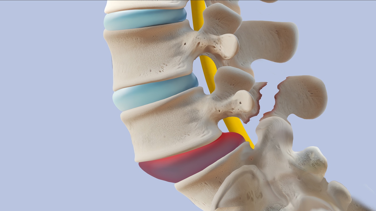
The spinal joint is composed of three parts: the disc in the front and two paired structures in the back called the facets. A pars defect is a gap in the pars interarticularis, a small bridge of bone that connects the vertebra above to the facet joint in the back. Although there are different reasons why pars defects occur, most of them are a result of stress fractures. Doctors often use the medical term spondylolysis to describe this fracture.
Unlike a broken bone caused by a major accident, a pars defect usually develops over time. Small amounts of repeated stress gradually weaken the bone until a tiny crack forms. Because the injury develops slowly, many people cannot remember one specific event that caused their symptoms.
The fracture most often occurs in the L5 vertebra, which sits at the bottom of the lumbar spine just above the sacrum. This area supports much of your body weight and absorbs force whenever you bend, twist, jump, or lift.
A pars defect can affect one side of the vertebra (unilateral) or both sides (bilateral). Fractures on both sides increase the risk of spondylolisthesis, where one vertebra slips forward over another.
Many people never experience symptoms. In fact, some pars defects are discovered incidentally during X-rays or MRI scans performed for unrelated back problems. Others develop persistent low back pain that gradually becomes more noticeable during sports, exercise, or prolonged standing.
Fortunately, most pars defects respond well to conservative treatment. Early diagnosis and proper management often allow patients to return to normal activities without surgery.
Understanding the Lumbar Spine
To understand a pars defect, it helps to know a little about how the lumbar spine works.
Your lumbar spine consists of five vertebrae, labeled L1 through L5. Between each vertebra sits a spinal disc that acts like a cushion, helping absorb shock during everyday movement.
The vertebrae are connected by small joints in the back called facet joints, which allow your back to bend, twist, and rotate while keeping the spine stable. The pars interarticularis is a narrow bridge of bone that connects these joints.
Although the pars is small, it plays an important role in supporting your spine. Every time you lean backward, rotate your torso, jump, or land from a height, force travels through this section of bone.
The L5 vertebra carries the most stress because it sits at the bottom of the lumbar spine. Over time, repeated stress can overwhelm the bone’s natural ability to repair itself, leading to a stress fracture.
For this reason, pars defects are more common in athletes than in less active people.
What Causes a Pars Defect?
Most pars defects develop because of repetitive stress, not a single traumatic injury.
Each time you bend backward or rotate your spine, the pars interarticularis absorbs force. Normally, healthy bone repairs this everyday wear and tear. However, when the stress becomes greater than the bone can repair, tiny cracks begin to develop. Eventually, those cracks can become a complete stress fracture.
Several factors increase the likelihood of developing a pars defect.
Repetitive Back Extension
Repeatedly arching the back places the greatest amount of stress on the pars interarticularis.
Sports that involve frequent back extension include:
- Gymnastics
- Diving
- Cheerleading
- Dance
- Volleyball
- Figure skating
Repeating these movements over time increases the risk of a stress fracture.
High-Impact Sports
Football, wrestling, weightlifting, rowing, and other high-impact activities place significant stress on the lower back.
Twisting, lifting, and repeated impact can also increase stress on the lower back.
Rapid Growth During Adolescence
Teenagers experience rapid growth while participating in competitive athletics. Sometimes muscles become stronger faster than bones adapt, making the developing spine more vulnerable to stress injuries. This is one reason pars defects are diagnosed most often in adolescents and young adults.
Genetics
Some people naturally have thinner pars bones or slight differences in the way their vertebrae develop. These inherited traits may increase the likelihood of developing a stress fracture when combined with repetitive physical activity.
There is also a developmental form of pars defect where, it is assumed, the pars simply does not form in utero.
Thus a family history of pars defects may increase your risk.
Overtraining
Too much training without enough rest can keep the bone from healing. Young athletes who play sports year-round have less time to recover, increasing their risk of a pars defect.
It's time to get back
to doing what you love.
Symptoms of a Pars Defect
When symptoms do appear, they usually develop slowly over time rather than after one specific injury. The pain often becomes more noticeable during sports or other activities that place repeated stress on the lower back.
Understanding the common symptoms can help you recognize when it’s time to seek medical evaluation.
Lower Back Pain
The most common symptom of a pars defect is a dull, aching pain in the lower back.
Unlike sore muscles, this pain often returns during activities like bending, twisting, jumping, or lifting. Many people notice that the pain:
- Gets worse during sports or exercise
- Increases after standing or walking for long periods
- Improves with rest
- Feels better after sleeping overnight
Pain usually stays in the lower back, but the severity can vary from person to person.
Muscle Tightness
Your muscles may tighten to protect the injured area.
Many patients develop:
- Tight hamstrings
- Stiffness in the lower back
- Reduced flexibility
- Difficulty bending forward
These changes can make everyday activities feel uncomfortable, even when the fracture itself is relatively small.
Pain During Back Extension
Pain that becomes worse when leaning backward is one of the most recognizable signs of a pars defect.
Movements that commonly trigger symptoms include:
- Performing a backbend
- Serving a volleyball
- Blocking during football
- Gymnastics tumbling
- Diving
- Certain dance movements
Because these activities repeatedly stress the pars interarticularis, symptoms often improve after taking a break from sports.
Can a Pars Defect Cause Nerve Symptoms?
Most pars defects do not compress nearby nerves.
If the fracture allows the vertebra to slip forward (spondylolisthesis), it may press on nearby nerves.
Possible nerve symptoms include:
- Pain that travels down the leg
- Numbness or tingling
- Muscle weakness
- Difficulty walking
Loss of bladder or bowel control is not a typical symptom of a pars defect. If it occurs, seek emergency medical attention immediately.
How Is a Pars Defect Diagnosed?
Diagnosing a pars defect begins with a conversation about your symptoms, activity level, and medical history. Your spine specialist will then perform a physical examination and order imaging tests to confirm the diagnosis.
Physical Examination
During your exam, your doctor will ask questions about:
- When your pain started
- Which activities make it worse
- Previous sports injuries
- Your overall activity level
They’ll also evaluate your:
- Range of motion
- Muscle strength
- Reflexes
- Walking pattern
- Flexibility
One common exam finding is pain that increases when standing on one leg while gently leaning backward. Although this test alone cannot diagnose a pars defect, it helps your doctor determine whether additional imaging is needed.
X-Rays
X-rays are usually the first imaging test ordered.
They allow doctors to look for fractures, changes in spinal alignment, or signs of vertebral slippage that are indicative of instability. While standard X-rays identify many pars defects, early stress fractures may not always be visible.
Your doctor may recommend additional imaging if symptoms strongly suggest a pars defect despite normal X-ray findings.
CT Scan
CT scans provide highly detailed images of bone.
They help confirm:
- Whether or not a fracture is present
- Whether one or both sides are affected
- Whether or not the fracture has a chance to heal without surgery
Doctors often use CT scans when they need a closer look at the fracture.
MRI
MRI is especially valuable because it can detect stress reactions before a complete fracture develops.
MRI is best at showing
- Bone swelling
- Soft tissues issues like herniated disc
- Whether there is nerve compression
- Other causes of back pain
For younger athletes, MRI offers another advantage because it does not expose patients to radiation.
Pars Defect vs. Spondylolysis vs. Spondylolisthesis
These terms are often used together (and sound a lot alike), which makes it easy to confuse them.
Although they are closely related, they describe different spine conditions.
Pars Defect
A pars defect is the actual stress fracture in the pars interarticularis.
Spondylolysis
Spondylolysis is the medical name for a pars defect. The two terms mean the same thing.
Spondylolisthesis
Spondylolisthesis occurs when a vertebra slips forward in relationship to the vertebra below it.
Not every pars defect leads to spondylolisthesis. Many patients never develop any vertebral slippage, especially when the fracture is diagnosed early and treated appropriately. Similarly, many patients with spondylolisthesis do not have pars defects.
However, bilateral pars defects increase the likelihood of instability because the spinal joint between two vertebra has been weakened.
If you have pars defects, your spine specialist will monitor for progression of slippage during follow-up visits and imaging studies.
Surgical Treatment for a Pars Defect
Surgery may be considered if:
- Pain continues after several months of nonsurgical treatment
- The fracture causes significant spinal instability
- Spondylolisthesis develops
- Nerve compression leads to weakness, numbness, or persistent leg pain
- These symptoms significantly interfere with work, sports, or daily life
Your spine surgeon will carefully review your symptoms, physical examination, and imaging before recommending an operation.
Direct Pars Repair.
This procedure repairs the fracture itself with the help of small screws or wires. This option is only available to young patients with no vertebral slippage. Most of these patients are the relatively rare individuals in their teens or early twenties whose back pain does not resolve when they are first diagnosed. The advantages of a direct pars repair over the alternative surgery, a spinal fusion, are that it is a smaller operation and that it maintains normal spine motion.
Minimally Invasive Spinal Fusion
If the fracture cannot be repaired or significant instability has developed, spinal fusion is only other option. During a spinal fusion, the two vertebrae are permanently joined to form a single bone. The surgery requires using bone grafts and larger metal implants.
Although a larger surgery than a direct pars repair, it a procedure that has seen many advances over the last 25 years. Many surgeons now perform lumbar fusion using minimally invasive techniques. Compared with traditional open surgery, these procedures often involve:
- Smaller incisions
- Less muscle damage
- Reduced blood loss
- Shorter hospital stays
- Faster early recovery
Although fusion eliminate movement at one spinal level, this change is not functionally significant, and the surgery is highly successful for this condition.
Nonsurgical Treatment for a Pars Defect
Most people with a pars defect improve without surgery. Conservative treatment focuses on relieving pain, allowing the stress fracture to heal, and preventing additional strain on the spine.
Your treatment plan will depend on your age, activity level, symptoms, and whether the fracture is new or has been present for some time.
Activity Modification
The first step is reducing activities that place repeated stress on the injured bone.
This doesn’t mean complete bed rest. Instead, your doctor may recommend avoiding activities that involve:
- Repeated back extension
- Heavy lifting
- Jumping
- Twisting
- High-impact sports
For young athletes, this often means taking a temporary break from competition while the fracture heals and a period of bracing. Walking, swimming, and stationary biking help maintain strength without putting too much stress on the spine.
As your symptoms improve, you’ll gradually return to your normal activities under your doctor’s guidance.
Physical Therapy
Physical therapy is one of the most important parts of treatment.
Rather than simply treating pain, physical therapy addresses the underlying movement patterns that contributed to the injury.
Your therapist may focus on:
- Strengthening your core muscles
- Improving hip strength
- Increasing flexibility
- Correcting posture
- Teaching proper lifting and movement techniques
A stronger core helps stabilize the spine and reduces stress on the pars interarticularis during everyday activities and sports. Physical therapy also helps lower the risk of future injuries after you return to normal activity.
Bracing
Some patients, particularly teenagers with newly diagnosed fractures, may benefit from wearing a back brace.
The brace helps limit movement while the bone heals.
Bracing is not necessary for everyone. Your spine specialist will determine whether it is appropriate based on your age, symptoms, activity level, and imaging results.
If prescribed, the brace is typically worn for several weeks while healing is monitored.
Medications
Medication does not heal the fracture itself, but it can help manage pain while the bone recovers.
Your doctor may recommend:
- Acetaminophen
- Nonsteroidal anti-inflammatory drugs (NSAIDs)
- Prescription pain medication for severe discomfort
Medication is usually combined with activity modification and physical therapy rather than used as a standalone treatment.
Steroid Injections
Most pars defects do not require injections.
If inflammation continues to cause pain, your doctor may recommend a steroid injection. These injections reduce inflammation and can make it easier to participate in physical therapy while recovery continues.
How Long Does Conservative Treatment Take?
Healing takes time. Many patients notice improvement within several weeks, but complete recovery often requires three to six months.
Your doctor will monitor healing and let you know when you can safely return to sports.
Recovery After Surgery
Recovery varies depending on the severity of the fracture and the kind of surgery you have had, whether you have had a direct pars repair, a minimally invasive fusion or a traditional “open” fusion. All surgeries, however, require new bone to grow. Because of this, a period of at least six months to a year is required before resuming your usual activities.
Early Recovery
Except for very unusual situations, there is no “bed rest.” Walking is encouraged, within limits. Lifting is restricted as are activities that involve twisting and bending. Some surgeons brace their patients after surgery. After a period of serval weeks to several months, you activity with be liberalized and you will be referred for physical therapy to work on core strengthening and stretching.
Returning to Sports
Returning to sports happens gradually and only when your surgeon is confident that sufficient bone healing has occurred.
Rather than jumping back into full competition, most athletes progress through several stages:
- Pain-free daily activities
- Light cardiovascular exercise
- Strengthening and flexibility training
- Sport-specific drills
- Full practice
- Competition
Returning too quickly increases the risk that the bone will fail to heal.
Long-Term Outlook
The outlook for most patients with a pars defect is excellent.
Most people return to work, school, and sports without significant ongoing pain. Learning proper body mechanics, maintaining core strength, and avoiding overtraining can help reduce the risk of future back injuries.
When to See a Spine Specialist
Many cases of low back pain improve with rest and conservative care. See a spine specialist if back pain lasts more than a few weeks or interferes with daily activities.
Schedule an appointment with a spine specialist if you experience:
- Low back pain that does not improve with time and rest
- Pain that repeatedly returns during sports or exercise
- Pain, numbness, tingling, or weakness in your legs
- Difficulty standing, walking, sleeping or participating in normal activities
Early evaluation is especially important for young athletes. Finding a pars defect early will direct the appropriate treatment regimen and may help prevent spinal instability and the need for surgery.
Seek emergency medical care if you suddenly lose bladder or bowel control or develop leg weakness. These symptoms may indicate a spinal emergency that requires prompt treatment.
Personalized Pars Defect Treatment at Neurosurgeons of New Jersey
At Neurosurgeons of New Jersey, our spine surgeons understand that every patient has different goals. Every patient has different goals, whether returning to competitive sports or staying active without pain.
That’s why we take a personalized, conservative-first approach to care. Your evaluation begins with a thorough review of your symptoms, medical history, physical examination, and imaging studies. We explain your diagnosis in clear language, answer your questions, and develop a treatment plan based on your lifestyle and long-term goals.
For most patients, treatment focuses on activity modification, physical therapy, and other nonsurgical options. If you need surgery, we offer advanced techniques, including minimally invasive procedures when appropriate.
Whether your symptoms began during sports or developed over time, our team can help you understand your treatment options.
Frequently Asked Questions
Can a Pars Defect Heal on Its Own?
Many pars defects improve with rest, activity modification, and physical therapy, especially when diagnosed early. Younger patients often have the best chance of healing because their bones are still developing. Many people become pain-free even if the fracture does not completely heal.
Can Adults Develop a Pars Defect?
Yes. Although pars fractures most commonly occur in teenagers and young athletes, adults can also be diagnosed with the condition. Rarely, adults develop a new fracture, but this usually requires a significant trauma. Most adults who receive a new diagnosis of pars defect have, in fact, an older injury went undiagnosed because it just never caused significant symptoms before.
What Happens if a Pars Defect Is Left Untreated?
Some pars defects never cause problems. However, continuing high-impact activities despite ongoing pain may increase the risk of the fracture worsening or developing into spondylolisthesis. Early evaluation and treatment can help prevent complications and support a faster recovery.

About Dr. Alfred T. Ogden
Dr. Alfred T. Ogden is an accomplished neurosurgeon in North Jersey and is a proud member of Neurosurgeons of New Jersey, practicing out of their Ridgewood office conveniently located on East Ridgewood Avenue. Dr. Ogden is internationally recognized as a leader in minimally invasive spine surgery.